A Shared Resource Fee-for-Service Laboratory
A Shared Resource Fee-for-Service Laboratory
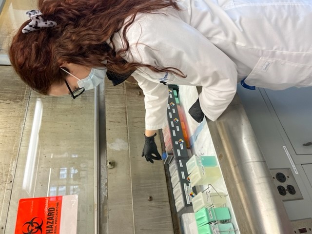
The Research Histology Laboratory is an integral and essential component of the Department of Pathology. It functions as the histology laboratory of the BioRepository & Precision Pathology Center and assists in the facilitation of both clinical trials and research requests. The laboratory also accepts requests directly from Duke investigators working with either human or animal tissue samples. The Research Histology Laboratory provides routine and specialized histological services, as well as integrated support with the other BRPC labs for immunohistochemical staining, nucleic acid extractions, whole slide imaging, tissue microarray creation, and digital spatial profiling. The staff can also provide guidance with respect to the proper collection and fixation of the tissue, and staining procedures.
Research Program Leader: Kate Frankey MHS, 919-684-6928
Director: Shannon J. McCall, MD
Staff:
Emily Knutson, AS, HT(ASCP), Team Lead
Kalpana Kodi, M.A, HT, HTL (ASCP)
Lab Telephone: 919-684-6209
Lab Shared Inbox: path-RHL@duke.edu
REQUESTS FOR SERVICE
Hours of Operation: Monday-Friday, 7:30AM-5:30PM
Designated drop off and pick up times: 9:30-11:00AM
Main Lab Space: Duke South Green Zone, Room 254
Please use this link to fill out a request https://redcap.duke.edu/redcap/surveys/?s=R7PX4CYTLWYJKRMR and email path-rhl@duke.edu with any questions.
SERVICES OFFERED
- Routine paraffin processing, embedding, and sectioning
- Paraffin processing, embedding, and sectioning from cells
- Hematoxylin and Eosin (H&E) Staining on FFPE and OCT samples
- Frozen Sectioning
- Special staining
SPECIAL STAINS
Stains are validated on FFPE samples only, unless noted otherwise. Other stains may be available. Please contact lab.
- Masson Trichrome
INSTRUMENTATION
- Sakura Tissue Tek VIP 5 tissue processors
- Sakura Tissue Tek E-300 processor
- Sakura Tissue Tek 5 tissue embedding consoles
- Leica RM2235 manual microtome
- Thermo Scientific/Epredia Microm HM355S automated microtomes
- Thermo Scientific CryoStar NX70 cryostat
- Leica Autostainer XL H&E Stainer
FAQs
Turnaround time varies depending on the project, specific services requested, and the number of samples involved in the project.
Yes – please clearly state on your request form that you have provided your own and label the containers to make sure they are used and returned to you.
Yes – please clearly state on your request form that you would like them returned and label the containers to make sure they are returned to you. If we do not have an indication that your container should be saved, it may be disposed of.
Yes – we can only ensure your samples are both returned to you and handled per your specifications if we can match them with a list. Providing a typed list also ensures the lab can accurately label any slides, as handwriting on blocks often smears during processing.
No – our lab is open daily from 9:30-11AM. Please feel free to stop in during these hours and someone will be available to assist you. Staff may be working in other lab spaces outside of these times, so please contact us if you need to make other arrangements.
Ask staff about current turnaround time (this depends on project size, complexity, current staffing, and other pending projects) – a rush may not be necessary for the project to be completed by your deadline.
We used charged slides manufactured by Cancer Diagnostics for the majority of our output. We also stock FisherBrand SuperFrost (recommended for IHC work), Cancer Diagnostics uncharged, and FisherBrand ProbeOn Plus slides, which can also be used upon request. If you have your own slides that you would like the lab to use, those may be provided during project drop off.
Our molecular precautions procedure requires our technicians to spray down all equipment and tools with an RNAse inhibiting spray, use molecular grade bottled water in waterbaths, wear gloves, and change microtome blades between individual samples. We always use this procedure when alerted that samples will be used for RNA extraction, and strongly encourage it for DNA extraction as well. This procedure is not necessary for standard staining or immunohistochemistry.
Yes – please contact Dr. Zuowei Su directly for any questions relating to IHC antibodies and work ups.
Yes – please let us know your samples will need extraction so we can coordinate with Dr. Xufeng Chen, who provides this service.
Yes – please let us know which slides need to be imaged and we will do so after staining. Alternatively, slides stained prior may also be sent for imaging only.